Life's hardships rewire the brain: Study pinpoints neural changes from adversity
In a recent study published in JAMA Network Open, researchers assess the link between adverse life experiences and changes in brain reactivity using the multilevel kernel density analysis (MKDA) method on task-based functional magnetic resonance imaging (fMRI) studies.
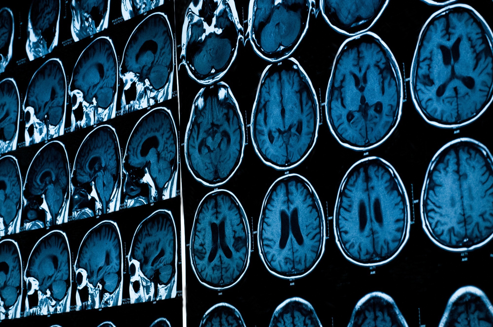
 Study: Adverse Life Experiences and Brain Function: A Meta-Analysis of Functional Magnetic Resonance Imaging Findings. Image Credit: Tushchakorn / Shutterstock.com
Background
Negative life experiences can alter brain functions, thereby increasing the risk of mental illnesses. The main brain regions affected include the prefrontal cortex (PFC), amygdala, and hippocampus.
While animal studies confirm this, human data is variable due to differences in defining adversity, measuring its impact, and diverse study methods. Variability also arises from the use of different image acquisition and analysis techniques.
A meta-analysis using the MKDA method, which accounts for these variations, provided more reliable insights than the activation likelihood estimation (ALE) method. However, given the inconsistencies in human studies on brain responses to adversity, further research is essential to understand long-term neuroplastic changes from adverse experiences.
About the study
The present study followed the Preferred Reporting Items for Systematic Reviews and Meta-Analyses (PRISMA) reporting guidelines. Comprehensive literature searches were performed across databases, including PsycINFO, Medline, EMBASE, and Web of Science, until May 2022. Additional searches in the Brainmap database and gray literature were also performed.
The search combined terms related to trauma, adversity, neuroimaging, and various cognitive processes. Articles were selected based on specific criteria, which led to the exclusion of conference abstracts, books, and certain other types of publications.
From the initial 2,016 abstracts identified, 336 met the criteria for a more in-depth review. Two reviewers assessed these articles, and a third reviewer resolved any discrepancies.
Brain activation coordinate data were precisely extracted and verified. To elucidate the diverse definitions of adversity across studies, these data were categorized based on criteria like threat or deprivation and by adversity severity.
For statistical analysis, the researchers extracted activation coordinates and grouped them based on task type and participant groups. The MKDA method was used to determine whether activations were consistent across studies.
Simulations were used to verify the authenticity of the findings. The data analysis was conducted between August and November 2022 using specialized software tools.
Study findings
In the comprehensive analysis of 83 studies comprising 5,242 participants, significant variations in blood-oxygen-level-dependent (BOLD) responses were observed in relation to adversity exposure. When the data from 67 studies was examined, those exposed to adversity exhibited enhanced right amygdala responses as compared to their counterparts. Comparatively, 47 other studies showed that the adversity group displayed consistently diminished responses in the medial frontal gyrus.
Of the 50 studies on emotion processing, the adversity-exposed group exhibited heightened amygdala activity and reduced superior frontal gyrus activity. In 11 studies focused on inhibitory control, those who experienced adversity exhibited increased activity in the claustrum, anterior cingulate cortex, and insula. No difference was observed in studies about memory or reward-processing tasks.
When examining threats as adversity, there was amplified BOLD response in the superior temporal gyrus and decreased medial frontal gyrus activity for the adversity group. This pattern persisted across different task domains.
When mixed types of adversities were studied, individuals exposed to these mixed adversities exhibited heightened activity across all domains in the right amygdala, precuneus, and superior frontal gyrus. In studies focusing solely on deprivation-type adversities, no significant results were reported, thus making definitive conclusions challenging.
Individuals exposed to trauma-type adversities exhibited significantly greater bilateral amygdala activation and reduced activity in areas like the medial frontal gyrus and anterior cingulate cortex. Meanwhile, moderate adversities were not associated with any significant associations.
The association between trauma and psychopathological conditions like post-traumatic stress disorder (PTSD) was also examined. To this end, individuals diagnosed with PTSD exhibited significantly greater left amygdala activation but diminished activity in regions like the hippocampus, orbitofrontal cortex, and insula.
The study also considered developmental stages by categorizing participants into adults, adolescents, and children. Adult data, which were extracted from 34 studies, revealed that adversity exposure during adulthood was associated with increased right amygdala activation but decreased activity in the middle frontal gyrus.
Conclusions
Individuals with a history of severe adversity have heightened amygdala responses and reduced PFC reactions to psychological challenges. MKDA of 83 studies showed potential PFC impairment in regulating amygdala activity.
The amygdala, which is essential for threat detection, exhibited increased activity as a PTSD biomarker. People with PTSD or those who experienced severe adversities exhibited more robust amygdala and weaker PFC responses across tasks.
Specific adversities increased insula and anterior cingulate cortex (ACC) activity. Traumatic adversities particularly enhanced amygdala activity.
Notably, individuals with PTSD exhibited significant left amygdala activity, which contrasts with some earlier studies, and decreased activity in other brain regions.
- Hosseini-Kamkar, N., Farahani, M. V., Nikolic, M., et al. (2023). Adverse Life Experiences and Brain Function: A Meta-Analysis of Functional Magnetic Resonance Imaging Findings. JAMA Network Open. doi:10.1001/jamanetworkopen.2023.40018
Posted in: Medical Science News | Medical Research News | Medical Condition News | Healthcare News
Tags: Adolescents, Amygdala, Biomarker, Blood, Brain, Children, Cortex, Hippocampus, Imaging, Magnetic Resonance Imaging, Neuroimaging, Oxygen, Post-Traumatic Stress Disorder, Research, Software, Stress, Trauma

Written by
Vijay Kumar Malesu
Vijay holds a Ph.D. in Biotechnology and possesses a deep passion for microbiology. His academic journey has allowed him to delve deeper into understanding the intricate world of microorganisms. Through his research and studies, he has gained expertise in various aspects of microbiology, which includes microbial genetics, microbial physiology, and microbial ecology. Vijay has six years of scientific research experience at renowned research institutes such as the Indian Council for Agricultural Research and KIIT University. He has worked on diverse projects in microbiology, biopolymers, and drug delivery. His contributions to these areas have provided him with a comprehensive understanding of the subject matter and the ability to tackle complex research challenges.