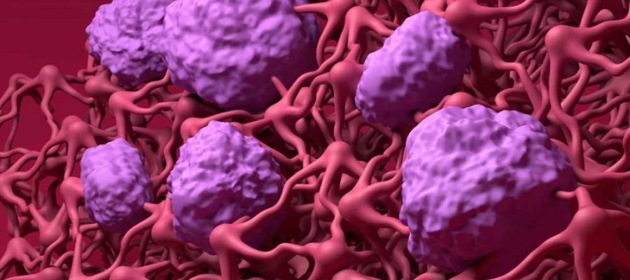
In a recent study published in the journal Nature Communications, researcher discovered that CD8+ T-cells are detained at the blood-brain barrier (BBB), wherein they contribute to its breakdown and subsequent infiltration of inflammatory cells into the brain.
 Study: Antigen recognition detains CD8+ T cells at the blood-brain barrier and contributes to its breakdown. Image Credit: Nemes Laszlo / Shutterstock.com
Background
Multiple sclerosis (MS) is a disease of the central nervous system (CNS) that causes inflammation and demyelination. Moreover, MS is characterized by the breakdown of the BBB and infiltration of immune cells.
New findings suggest that CD8+ T-cells have unique ways of crossing the BBB that differ from CD4+ T-cells. CD4+ T-cell migration across the BBB does not require Ag-specific mechanisms; however, the transfer of CNS autoantigen-specific CD8+ T-cells across the BBB appears to be dependent on the luminal expression of major histocompatibility complex (MHC) class I. This suggests that brain endothelial cells carry CNS antigens from the abluminal side for processing and subsequent presentation to circulating CD8+ T-cells on the luminal side.
About the study
The impact of brain endothelial cell Ag presentation on CD8+ T-cell effector functions was determined using the induction of effector molecule expression in OT-I cells following co-incubation with tumor necrosis factor α (TNF-α)/interferon γ (IFN-γ) stimulated pMBMECs that were either pulsed with SIINFEKL or vesicular stomatitis virus (VSV) peptide or loaded with ovalbumin (OVA).
The interaction between naïve OT-I cells and unpulsed or SIINFEKL-pulsed primary brain microvascular endothelial cells (pMBMECs) over time was observed through live cell imaging.
The researchers also explored whether pMBMECs could uptake and process exogenous antigens from their abluminal side and present these antigens on their luminal surface to naïve CD8+ T-cells. This was performed by growing pMBMECs on filter inserts, stimulating these cells with luminal TNF-α/IFN-γ, and subsequently subjecting pMBMECs to increasing proportions of OVA or SIINFEKL from the abluminal compartment and naïve OT-I cells on the luminal surface.
Study findings
Non-stimulated pMBMECs exhibited low cell surface staining for MHC class I, while no staining was observed for CD80, CD86, or programmed death-ligand 1 (PD-L1). After TNF-α/IFN-γ stimulation for 24 and 48 hours, MHC class I and PD-L1 staining increased in pMBMECs, whereas CD80 and CD86 levels were not affected.
After 48 hours of co-incubation with SIINFEKL-pulsed and OVA-loaded pMBMECs, OT-I cells exhibited upregulation of IFN-γ and granzyme B (GrB). Additionally, after 72 hours, OT-I cells co-incubated with SIINFEKL-pulsed and OVA-loaded pMBMECs exhibited an upregulation of TNF-α, GrB, perforin, lysosome-associated membrane glycoprotein (LAMP)-1, and fas ligand (FasL); however, this was not observed in VSV-peptide pulsed and unpulsed pMBMECs.
Exposure of abluminal pMBMECs to SIINFEKL or OVA protein resulted in the activation of naïve OT-I cells, as demonstrated by the increased expression of CD25, CD69, and CD44, and shedding of CD62L. Comparatively, OT-I cells that were cocultured with pMBMECs without added abluminal Ag remained in a naïve state.
Activation of OT-I cells resulted in their proliferation and differentiation into effector cells, which caused disruption of the pMBMEC monolayers. The ability of pMBMECs to present exogenous Ag on their luminal surface through MHC class I molecules allows for the activation and differentiation of naïve CD8+ T-cells into effector CD8+ T-cells, which can lead to focal BBB breakdown.
The number of activated OT-I cells that were arrested on pMBMECs was seven times higher as compared to naïve OT-I cells. The initial arrest of activated OT-I cells was not influenced by the presence or absence of functional MHC class I or cognate Ag.
Effector OT-I cells crossed the WT or B2M–/– pMBMEC monolayer in the absence of SIINFEKL after probing or crawling. However, OT-I cells exhibited decreased crawling and increased probing behavior without diapedesis when exposed to SIINFEKL-pulsed WT pMBMECs, as opposed to the control group.
OT-I cells labeled with 5-chloromethyl fluorescein diacetate (CMFDA) and in vitro activated effector OT-I cells were compared with the luminal wall of spinal cord microvessels in ODC-OVA and control mice. To this end, OT-I cells in ODC-OVA mice had a considerably reduced crawling speed.
Conclusions
Brain endothelial cells can uptake, process, and present exogenous antigens to CD8+ T-cells in vitro and in vivo. Furthermore, presenting antigens on the BBB led to the arrest of CD8+ T-cells specific to those antigens, subsequently causing immune infiltration into the brain and apoptosis in brain endothelial cells.
The researchers also discovered that focal BBB breakdown was not related to the cytotoxic action of CD8+ T-cells. In ODC-OVA models, CNS-resident macrophages efficiently took up OVA, thereby resulting in limited antigen uptake and BBB presentation. Nevertheless, antigen presentation reduced the crawling speed of OVA-specific CD8+ T-cells in CNS microvessels.
The current study suggests that neuroinflammation can cause MHC class I-restricted Ag presentation at the BBB, which may lead to reduced CD8+ T-cell entry into the CNS. High-level Ag presentation at the BBB could lead to the focal breakdown of BBB through CD8+ T-cell activation.
- Aydin, S., Pareja, J., Schallenberg, V. M., et al. (2023). Antigen recognition detains CD8+ T cells at the blood-brain barrier and contributes to its breakdown. Nature Communications 14(1); 1-20. doi:10.1038/s41467-023-38703-2
Posted in: Molecular & Structural Biology | Medical Science News | Medical Research News
Tags: Antigen, Apoptosis, Blood, Brain, CD4, Cell, Cell Imaging, Cell Migration, Central Nervous System, Demyelination, Endothelial cell, Glycoprotein, Imaging, in vitro, in vivo, Inflammation, Interferon, Ligand, Live Cell Imaging, Membrane, Molecule, Multiple Sclerosis, Necrosis, Nervous System, PD-L1, Perforin, Proliferation, Protein, Sclerosis, Stomatitis, T-Cell, Tumor, Tumor Necrosis Factor, Virus

Written by
Bhavana Kunkalikar
Bhavana Kunkalikar is a medical writer based in Goa, India. Her academic background is in Pharmaceutical sciences and she holds a Bachelor's degree in Pharmacy. Her educational background allowed her to foster an interest in anatomical and physiological sciences. Her college project work based on ‘The manifestations and causes of sickle cell anemia’ formed the stepping stone to a life-long fascination with human pathophysiology.
Source: Read Full Article
