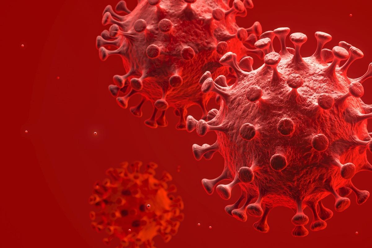
In a recent study posted to the bioRxiv* pre-print server, researchers investigated coronavirus disease 2019 (COVID-19)-triggered changes in the levels of cyclins and cyclin-dependent kinases (CDKs) inside the host cells.

Both cyclins and CDKs are key regulators of the host cell cycle. Many coronaviruses (CoVs), including severe acute respiratory syndrome coronavirus 2 (SARS-CoV-2), translocate and degrade cyclins inside the host cells for replicating efficiently.
A recent study investigating phosphoproteomic profiles of SARS-CoV-2-infected VERO E6 cells revealed that SARS-CoV-2 diminished CDK1 and CDK2 activities. In this way, viruses arrest the host cell cycle during the deoxyribonucleic acid (DNA) synthesis (S) phase and cell growth (G2) phase.
It is noteworthy that pathogenic viruses use host-cell synthesized nucleotides for their replication. Also, by arresting host cell cycle arrest, they increase DNA repair processes and replication proteins crucial for viral replication.
About the study
In the present study, researchers gathered extensive data on SARS-CoV-2 cell cycle changes using VERO and A549 alveolar type II (AT2) cells.
They used immunofluorescence to analyze the cellular distribution of cyclins D1, D3, and A2 in infected VERO AT2 cells. The fluorescence intensity reflected the nucleus and cytoplasm (N/C) ratio of cyclins in uninfected and infected cells. Further, the researchers measured D-cyclin levels in the infected cells in the absence or presence of protease inhibitors, carbobenzoxy-Leu-Leu-leucinal (MG-132) and Bortezomib.
The small interfering RNA (siRNA) reduces cyclin D and A2 levels by more than 80% at 48h post-transfection in A549 AT2 cells compared to non-targeted controls. The cells depleted of cyclins were exposed to Delta, Alpha, and wildtype SARS-CoV-2 variants multiple times to investigate the functional role of D-cyclins in SARS-CoV-2 pathogenesis.
The team used the fluorescence ubiquitination cell-cycle indicator (FUCCI) (or sensor) in VERO and A549 AT2 cells to examine the cell cycle arrest phenotype. Lastly, they used flow cytometry to visualize G1, early S, S, G2, and mitotic (M) phases.
Study findings
The study findings indicated that SARS-CoV-2 infection triggered the translocation of cyclin D1 and cyclin D3 from the host cell nucleus into cytoplasm for proteasome degradation and restored its assembly and release of newly produced virions.
Fucci sensor expression helped the researchers quantify the N/C ratio of D-cyclin staining in different cell cycle phases. The results showed that regardless of the cell cycle phase, cyclin D3 and cyclin D1 in SARS-CoV-2-infected cells were relocalized from nucleus to cytoplasm for degradation. Intriguingly, SARS-CoV-2 induced only cyclin D protein degradation. Accordingly, analysis of other cyclins in VERO AT2 cells did not show any changes to cyclins A, B, or E.
Cyclin A promotes S phase entry and mitosis, cyclin B accumulates in the cell growth (G2) phase, and cyclin E prevents entry into the S phase. Moreover, the specific cell cycle phases in which cells are arrested appear to be dependent on the cell type.
With diminished cyclin D1 and cyclin D3 levels, the host cell cycle arrest in the G1 phase, not in the S/G2 phase. In the absence of cyclin Ds, A549 AT2 cells were arrested in the early S phase and VERO AT2 cells in the S/G2 or M phase, indicating the role of other factors in cell cycle arrest during COVID-19.
Notably, cyclin D proteins always re-localized from nucleus to cytoplasm and degraded in all cell cycle phases during COVID-19. Conversely, the expression levels of CDKs did not change. The authors believed that the phosphorylation state of CDKs might have impacted cell cycle progression; however, the present study did not investigate this effect.
SARS-CoV-2 requires M, E, and nucleocapsid (N) proteins for virion assembly in the endoplasmic reticulum (ER)-Golgi intermediate compartment cisternae. The authors noted the role of cyclin D3 in SARS-CoV-2 spread and defective viral assembly. More importantly, it emerged as a new interactor with SARS-CoV-2 envelope (E) and even membrane (M) protein.
Cyclin D3 associates with SARS-CoV-2 M protein to impair SARS-CoV-2 assembly and spread. Conversely, host cells retain SARS-CoV-2 spike (S) protein in the presence of M and E; however, its translocation towards the membrane and ability to form syncytia diminishes due to cyclin D3. The data support the hypothesis that cyclin D3 is a restriction factor that impedes the role of M and E during SARS-CoV-2 assembly.
Conclusions
The study highlighted how cyclin D proteins interfere with SARS-CoV-2 E and M proteins to impair efficient viral assembly and replication. Therefore, SARS-CoV-2 has evolved strategies to degrade cyclin D3 for more efficient virion assembly and replication. This crucial phenomenon needs to be extensively investigated across all SARS-CoV-2 variants to develop a universal antiviral target against SARS-CoV-2.
*Important notice
bioRxiv publishes preliminary scientific reports that are not peer-reviewed and, therefore, should not be regarded as conclusive, guide clinical practice/health-related behavior, or treated as established information.
- Ravindra K Gupta, Petra Mlcochova. (2022). Cell cycle independent role of cyclin D3 in host restriction of SARS-CoV-2 infection. bioRxiv. doi: https://doi.org/10.1101/2022.05.07.491022 https://www.biorxiv.org/content/10.1101/2022.05.07.491022v1
Posted in: Medical Science News | Medical Research News | Disease/Infection News
Tags: Cell, Cell Cycle, Cell Nucleus, Coronavirus, covid-19, Cytometry, Cytoplasm, DNA, Flow Cytometry, Fluorescence, Membrane, Mitosis, Nucleotides, Phenotype, Phosphorylation, Protein, Respiratory, RNA, SARS, SARS-CoV-2, Severe Acute Respiratory, Severe Acute Respiratory Syndrome, siRNA, Syndrome, Transfection

Written by
Neha Mathur
Neha is a digital marketing professional based in Gurugram, India. She has a Master’s degree from the University of Rajasthan with a specialization in Biotechnology in 2008. She has experience in pre-clinical research as part of her research project in The Department of Toxicology at the prestigious Central Drug Research Institute (CDRI), Lucknow, India. She also holds a certification in C++ programming.
Source: Read Full Article
