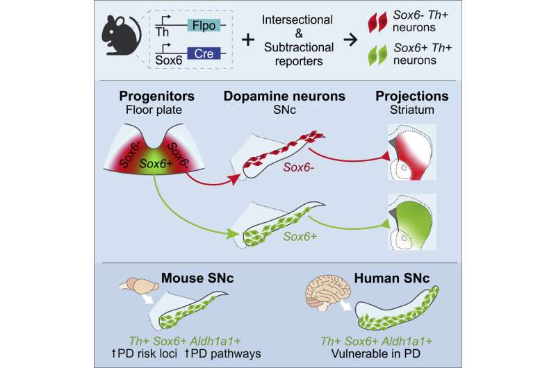
Northwestern investigators have discovered the molecular signature of a subset of dopaminergic neurons with an increased vulnerability to degeneration.
The findings shed light on the mechanisms at play within a small subsection of the brain’s substantia nigra, called the pars compacta, which contribute to the development of Parkinson’s disease and may help identify novel therapeutic targets, according to a study published in the journal Cell Reports.
“This provides a better understanding into the neuronal diversity within the substantia nigra and why some cohorts may be more vulnerable than others. In doing so, it might provide a better understanding of the degenerative underpinnings of Parkinson’s disease,” said Rajeshwar Awatramani, Ph.D., professor in the Ken and Ruth Davee Department of Neurology’s Division of Movement Disorders and senior author of the study.
Milagros Luppi, a student in the Driskill Graduate Program in Life Sciences (DGP), Maite Azcorra, a student in the Northwestern University Interdepartmental Neuroscience (NUIN) program, and Giuliana Caronia-Brown, Ph.D., research assistant professor of Pharmacology, were co-first authors of the study.
A prominent feature of Parkinson’s disease is the degeneration of the brain’s substantia nigra, a structure located in the midbrain containing dopaminergic neurons and which serves a critical role in reward response and movement. Parkinson’s disease is characterized by the selective loss of dopaminergic neurons in the substantia nigra’s ventral tier, while dopaminergic neurons in its dorsal tier are generally untouched. The molecular mechanisms that contribute to this disparity, however, have remained unknown.
For the current study, Awatramani and colleagues performed immunofluorescence staining on post-mortem samples of substantia nigra from patients with Parkinson’s disease and discovered that ventral dopaminergic neurons express different molecular signatures than dorsal dopaminergic neurons.
Specifically, ventral dopaminergic neurons express the transcription factor Sox6 and are enriched in mitochondrial pathways such as mitochondrial biogenesis and electron transport, which may increase their vulnerability to degeneration, according to Awatramani.
“Now, we have a molecular-genetic handle on this population that turns out to be more vulnerable,” said Awatramani, who is also a member of the Robert H. Lurie Comprehensive Cancer Center of Northwestern University.
Using mouse models, the investigators further demonstrated that Sox6-positive and -negative dopaminergic neurons project throughout different areas of the basal ganglia; Sox6-positive neurons projected to the dorsal striatum, and their activity in the dorsal most aspect of the striatum correlates with acceleration, and Sox6-negative neurons projected to the medial, ventral and caudal striatum and may respond to rewards.
Furthermore, the investigators found that dopamine progenitor cells are divided into Sox6-positive progenitor cells and Sox6-negative progenitor cells during early embryonic development.
“The difference between dorsal and ventral that manifests in symptoms of vulnerability in terms of function and molecular signature is encoded very early in the embryo,” Awatramani said.
Source: Read Full Article
